Page 1 of 1
ACTIVITY 21.3 378 Examining the Internal Anatomy of the Heart: The Heart Chambers The two atria are thin-walled chambers
Posted: Mon Jul 11, 2022 2:28 pm
by answerhappygod
- Activity 21 3 378 Examining The Internal Anatomy Of The Heart The Heart Chambers The Two Atria Are Thin Walled Chambers 1 (46.08 KiB) Viewed 90 times
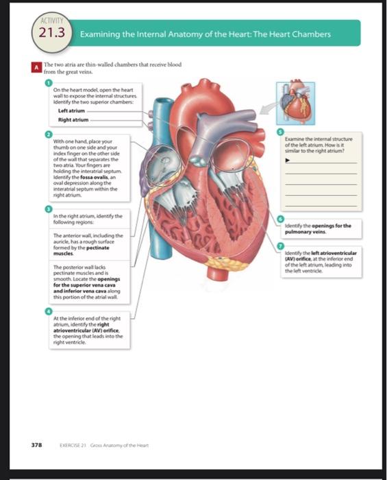
ACTIVITY 21.3 378 Examining the Internal Anatomy of the Heart: The Heart Chambers The two atria are thin-walled chambers that receive blood from the great veins. On the heart model open the heart wall to expose the internal structures identify the two superior chambers: Left atrium. Right atrium- With one hand, place your thumb on one side and your index finger on the other side of the wall that separates the two atria. Your fingers are holding the interatrial septum identify the fossa ovalis, an oval depression along the interatrial septum within the right atrium In the right atrium, identify the following regions The anterior wall, including the auricle, has a rough surface formed by the pectinate muscles The posterior wall lacks pectinate muscles and is smooth. Locate the openings for the superior vena cava and inferior vena cava along this portion of the atrial wall At the inferior end of the right atrium, identify the right atrioventricular (AV) orifice the opening that leads into the EXERCISE 21 Cross Anatomy of the Heart Examine the internal structure of the left atrium How is it similar to the right atrium? Identify the openings for the pulmonary veins. Identify the left atrioventricular (AV) orifice at the inferior end of the left atrium, leading into the left ventricle