Page 1 of 1
17 Superior or inferior vena cava valve valve trunk 2. In the chart shown in FIGURE 17.16, fill in the blood flow pathwa
Posted: Thu Jul 07, 2022 12:46 pm
by answerhappygod
- 17 Superior Or Inferior Vena Cava Valve Valve Trunk 2 In The Chart Shown In Figure 17 16 Fill In The Blood Flow Pathwa 1 (46.83 KiB) Viewed 31 times
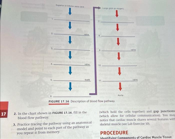
17 Superior or inferior vena cava valve valve trunk 2. In the chart shown in FIGURE 17.16, fill in the blood flow pathway. 10. FIGURE 17.16 Description of blood flow pathway. 3. Practice tracing the pathway using an anatomical model and point to each part of the pathway as you repeat it from memory. Lungs (pick up oxygen) Aorta valve valve (which hold the cells together) and gap junctions (which allow for cellular communication). You may notice that cardiac muscle shares several features with skeletal muscle (see Lab Exercise 10). PROCEDURE Identifying Components of Cardiac Muscle Tissue