Page 1 of 1
Relaxation weighted imaging sequences A region of the brain to be imaged contains areas corresponding to tumour, normal
Posted: Mon May 23, 2022 10:09 am
by answerhappygod
- Relaxation Weighted Imaging Sequences A Region Of The Brain To Be Imaged Contains Areas Corresponding To Tumour Normal 1 (45.23 KiB) Viewed 21 times
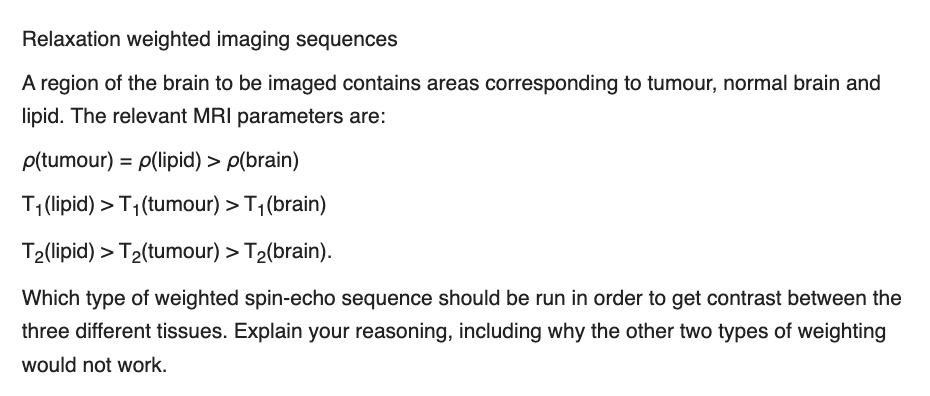
Relaxation weighted imaging sequences A region of the brain to be imaged contains areas corresponding to tumour, normal brain and lipid. The relevant MRI parameters are: p(tumour) = p(lipid) > p(brain) T(lipid) >T1(tumour) > T1(brain) T2(lipid) > T2(tumour) > T2(brain). Which type of weighted spin-echo sequence should be run in order to get contrast between the three different tissues. Explain your reasoning, including why the other two types of weighting would not work.