Page 1 of 1
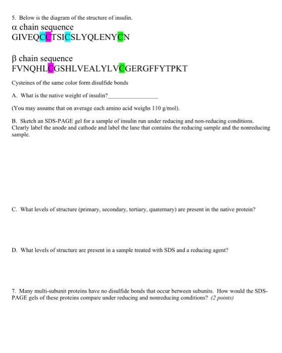
5. Below is the diagram of the structure of insulin. a chain sequence GIVEQCCTSICSLYQLENYON B chain sequence FVNQHLCGSHL
Posted: Wed May 18, 2022 10:27 am
by answerhappygod
- 5 Below Is The Diagram Of The Structure Of Insulin A Chain Sequence Giveqcctsicslyqlenyon B Chain Sequence Fvnqhlcgshl 1 (35.9 KiB) Viewed 78 times
5. Below is the diagram of the structure of insulin. a chain sequence GIVEQCCTSICSLYQLENYON B chain sequence FVNQHLCGSHLVEALYLVCGERGFFYTPKT Cysteines of the same color form disulfide bonds A. What is the native weight of insulin? (You may assume that on average each amino acid weighs 110 g/mol). B. Sketch an SDS-PAGE gel for a sample of insulin run under reducing and non-reducing conditions. Clearly label the anode and cathode and label the lane that contains the reducing sample and the nonreducing sample. C. What levels of structure (primary, secondary, tertiary, quaternary) are present in the native protein? D. What levels of structure are present in a sample treated with SDS and a reducing agent? 7. Many multi-subunit proteins have no disulfide bonds that occur between subunits. How would the SDS- PAGE gels of these proteins compare under reducing and nonreducing conditions? (points)