- Nervous Control Of Ventilation There Is A Negative Feedback Loop That Helps Control Ventilation The Stimulus Is When Th 1 (66.99 KiB) Viewed 42 times
- Nervous Control Of Ventilation There Is A Negative Feedback Loop That Helps Control Ventilation The Stimulus Is When Th 2 (15.99 KiB) Viewed 42 times
- Nervous Control Of Ventilation There Is A Negative Feedback Loop That Helps Control Ventilation The Stimulus Is When Th 3 (13.15 KiB) Viewed 42 times
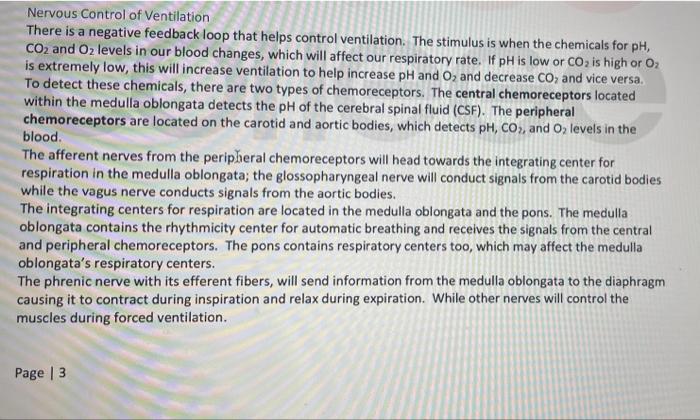
Nervous Control of Ventilation There is a negative feedback loop that helps control ventilation. The stimulus is when the chemicals for pH, CO2 and Oz levels in our blood changes, which will affect our respiratory rate. If pH is low or CO2 is high or 02 is extremely low, this will increase ventilation to help increase pH and Oz and decrease CO and vice versa. To detect these chemicals, there are two types of chemoreceptors. The central chemoreceptors located within the medulla oblongata detects the pH of the cerebral spinal fluid (CSF). The peripheral chemoreceptors are located on the carotid and aortic bodies, which detects pH, CO2, and O, levels in the blood. The afferent nerves from the peripheral chemoreceptors will head towards the integrating center for respiration in the medulla oblongata; the glossopharyngeal nerve will conduct signals from the carotid bodies while the vagus nerve conducts signals from the aortic bodies. The integrating centers for respiration are located in the medulla oblongata and the pons. The medulla oblongata contains the rhythmicity center for automatic breathing and receives the signals from the central and peripheral chemoreceptors. The pons contains respiratory centers too, which may affect the medulla oblongata's respiratory centers. The phrenic nerve with its efferent fibers, will send information from the medulla oblongata to the diaphragm causing it to contract during inspiration and relax during expiration. While other nerves will control the muscles during forced ventilation. Page 3
The effector during eupnea is only the diaphragm during inspiration and relaxation of the diaphragm during expiration, so no muscles contract during resting expiration. During forced inspiration, the external intercostals, scalenes, and sternocleidomastoid contract, while during forced expiration the abdominal muscles and internal intercostals contract. The response will be the opposite of the stimulus to maintain homeostasis, creating a negative feedback loop.
Draw a feedback loop for ventilation. Your choice as to what the stimulus is.