Page 1 of 1
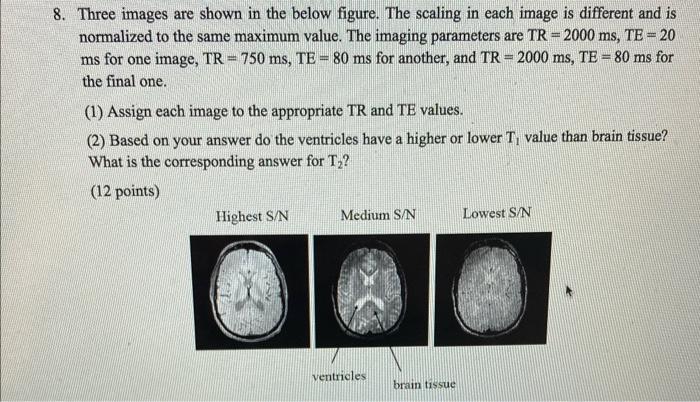
- 8. Three images are shown in the below figure. The scaling in each image is different and is normalized to the same ma
Posted: Tue May 17, 2022 3:41 pm
by answerhappygod
- 8 Three Images Are Shown In The Below Figure The Scaling In Each Image Is Different And Is Normalized To The Same Ma 1 (69.59 KiB) Viewed 90 times
- 8. Three images are shown in the below figure. The scaling in each image is different and is normalized to the same maximum value. The imaging parameters are TR = 2000 ms, TE = 20 ms for one image, TR = 750 ms, TE = 80 ms for another, and TR= 2000 ms, TE = 80 ms for the final one. (1) Assign each image to the appropriate TR and TE values. (2) Based on your answer do the ventricles have a higher or lower T, value than brain tissue? What is the corresponding answer for T,? (12 points) Highest S/N Medium S/N Lowest S/N ventricles brain tissue